|
|
|
|


|

|

|

|

|

|

|

|

|

|
|
|
|
|
|
|
|
|
|
|
|
|
|
|
|
|
Effect of Curvature on Cell Behavior
Michelle Gaines, Ya-Wen Chang, Savannah Carlsen, & Alberto Fernandez-Nieves
|
The properties of the extracellular microenvironment strongly influence epithelial cell behaviors such as proliferation, migration, differentiation and apoptosis. For tissue repair and embryogenesis, these behaviors are favorable, however in diseases such as cancer and organ fibrosis, collective epithelial cell growth is invasive and uncontrolled, and often leads to mortality. While the idea of directing cell behavior using geometrical cues has long been recognized, there is still a significant lack of understanding on how substrate curvature regulates cell behaviors, particularly those related to epithelial multi-cellular clusters. This gap in the knowledge must be closed, in order to collect the full perspective on how single-cell interfacial behavior directs collective cell behavior, which will be necessary to gain control over invasive cell growth in fibrosis and cancer.
Tissue Cell Behavior on Curved Substrates
Our approach is to study cell differentiation and proliferation on curved substrates at low cell density and as they approach jamming transition.
The unique geometry of a torus enables us to explore the effect of positive and negative gaussian curvature on cell behavior in one single structure.
We're interested in understanding how cells respond to the topological and geometric features of the environemnt collectively and as individuals.
In this work, we are by depositing MDCK II cells onto polyacrylamide hydrogel substrates that are both planar and torus-shaped.
We are using confocal microscopy and image analysis to quantify the local cell density of proliferating cells on the flat gels and the tori of different aspect ratios.
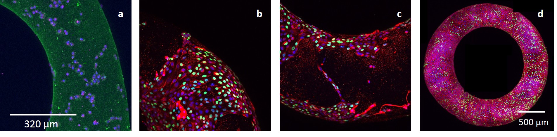
Polyacrylamide toroidal hydrogels are fabricated and seeded with MDCK epithelial cells.
Fig. 1: MDCK cells on toroidal hydrogel. 2D Max intensity projections of confocal microscopy z-stacks after (a) 2 hours, (b-c) 26 hours, and (d) 52 hours of incubation. |
Soft Condensed Matter Laboratory, School of Physics, Georgia Institute of Technology
770 State Street NW, Atlanta, GA, 30332-0430, USA
Phone: 404-385-3667 Fax: 404-894-9958
alberto.fernandez [at] physics.gatech.edu